
By Sanne Tol
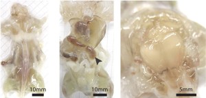
Is it Halloween yet? A picture for the strongly stomached: no it is not a gummy rat gone bad, it is an actual see-through rat. Scientists have developed a new technique of making rats transparent, allowing them to see more anatomical detail than ever before. These details are necessary for high quality 3D mapping of organs, which is important in finding details of structure-function relationships, which are currently unavailable for many organs across species.
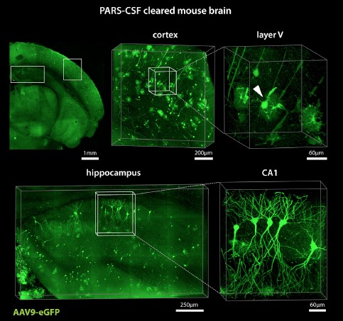
Research of making tissues clearer has been long going on, but (successful) attempts take months. A research team from California Institute of Technology, and David Geffen School of Medicine at UCLA, speeded things up by combining three techniques that are relatively easy and cost-effective. The introduced techniques are: PACT (passive clarity technique), a passive tissue clearing and immune-staining (meaning the staining of certain molecules concerned with the immune system) of intact organs; RIMS (refractive index matching solution), which is a mounting media for imaging thick tissue, such as organs, reserving fluorescent markers for months; and PARS (perfusion-assisted agent release in situ), the technique for clearing the entire body, and immunolabeling with endogenous fluorescence. The rats are treated with these technique by firstly pumping the necessary chemicals through the vessels, brain, and spinal cord to form a mesh to hold tissues in place, as it’s essential the organs should stay intact. Secondly, chemicals are pumped through to clear out fatty lipids that make organs opaque. After a few days most organs, excluding the brain, would turn clearer, and after two weeks all organs would turn clear.
After clearing out the rat, tissue sample can be mounted on the microscope, offering highly detailed photos of the fluorescently marked tissues. Specifically the detail of neurons is of interest, so says one of the researchers, V. Gradinaru: “We can obtain better maps for neuronal networks. A transparent whole organism is particularly useful to map the distribution and identity of peripheral nerves at their target organs.”
Researchers hope that these high quality mappings of the nervous system and their structure-function relationship, aid in the development of therapies for disorders such as migraine and epilepsy. In addition, the high quality mapping may also aid in cancer research, and virus research, allowing the researchers to follow processes on a level that has not yet been reached before.
By Sanne Tol, class of 2016, majoring in Biology, hometown: Bergen op Zoom
